Ap4C MV annulus diameter
Acquire an apical 4C view. Optimize the mitral annulus by adjusting the gains and focal zone to ensure adequate definition of the leaflet and their insertions into the annulus. Freeze and scroll to an early diastolic frame. Measure from where the anterior leaflet inserts into the annulus to the posterior leaflet insertion (using inner edge to inner edge technique).
AP4C TV annulus diameter
Acquire an apical 4C view. Optimize the tricuspid annulus by adjusting the gains and focal zone to ensure adequate definition of the leaflet and their insertions into the annulus. Freeze and scroll to an early diastolic frame. Measure from where the anterior leaflet inserts into the annulus to the septal leaflet insertion (using inner edge to inner edge technique).
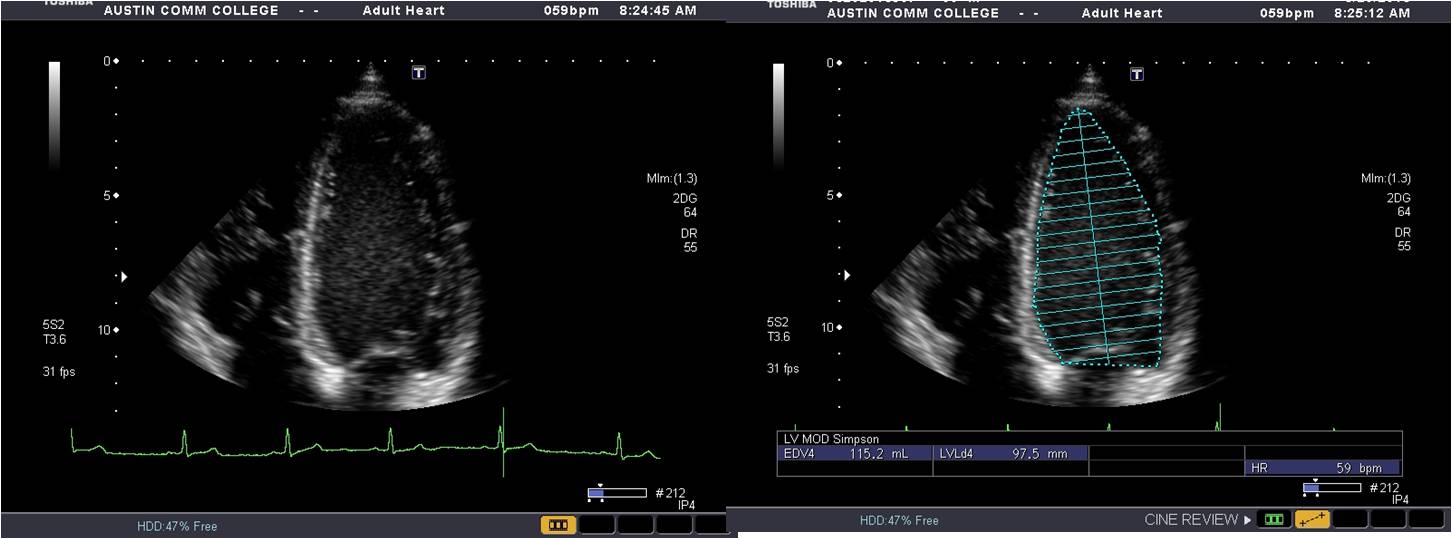
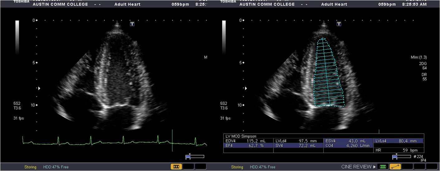
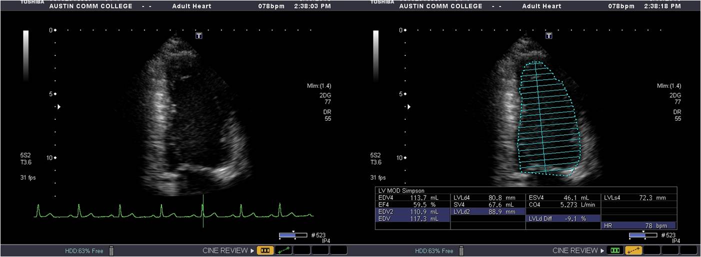
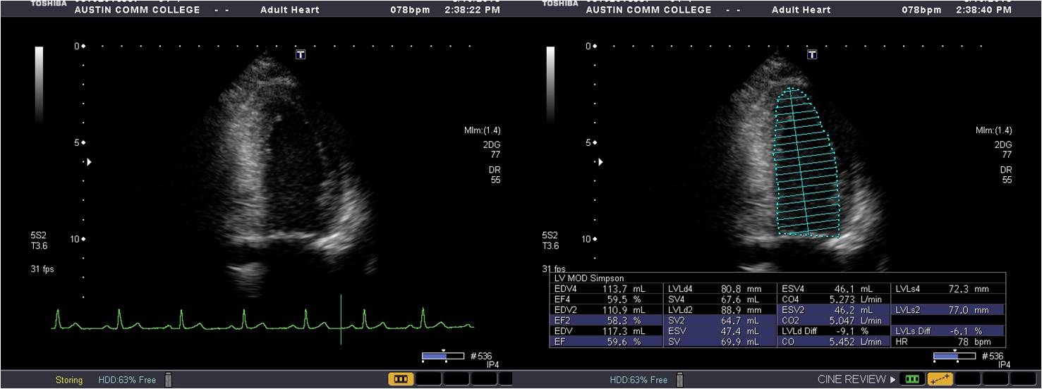
Ap4C LV volume @ end diastole (MOD)
Acquire an apical 4C view. Optimize the LV by decreasing the depth and adjusting the gains and focal zone to ensure adequate endocardial border definition. Freeze and scroll to an end-diastolic frame. Trace the borders and measure the length from the apex to the mitral valve annulus. Exclude the papillary muscles and trabeculations.
Ap4C LV volume @ end systole (MOD)
Acquire an apical 4C view. Optimize the LV by decreasing the depth and adjusting the gains and focal zone to ensure adequate endocardial border definition. Freeze and scroll to an end-systolic frame. Trace the borders and measure the length from the apex to the mitral valve annulus. Exclude the papillary muscles and trabeculations.
AP2C LV volume @ end diastole (MOD)
Acquire an apical 2C view. Optimize the LV by decreasing the depth and adjusting the gains and focal zone to ensure adequate endocardial border definition. Freeze and scroll to an end-diastolic frame. Trace the borders and measure the length from the apex to the mitral valve annulus. Exclude the papillary muscles and trabeculations.
AP2C LV volume @ end systole (MOD)
Acquire an apical 2C view. Optimize the LV by decreasing the depth and adjusting the gains and focal zone to ensure adequate endocardial border definition. Freeze and scroll to an end-systolic frame. Trace the borders and measure the length from the apex to the mitral valve annulus. Exclude the papillary muscles and trabeculations.
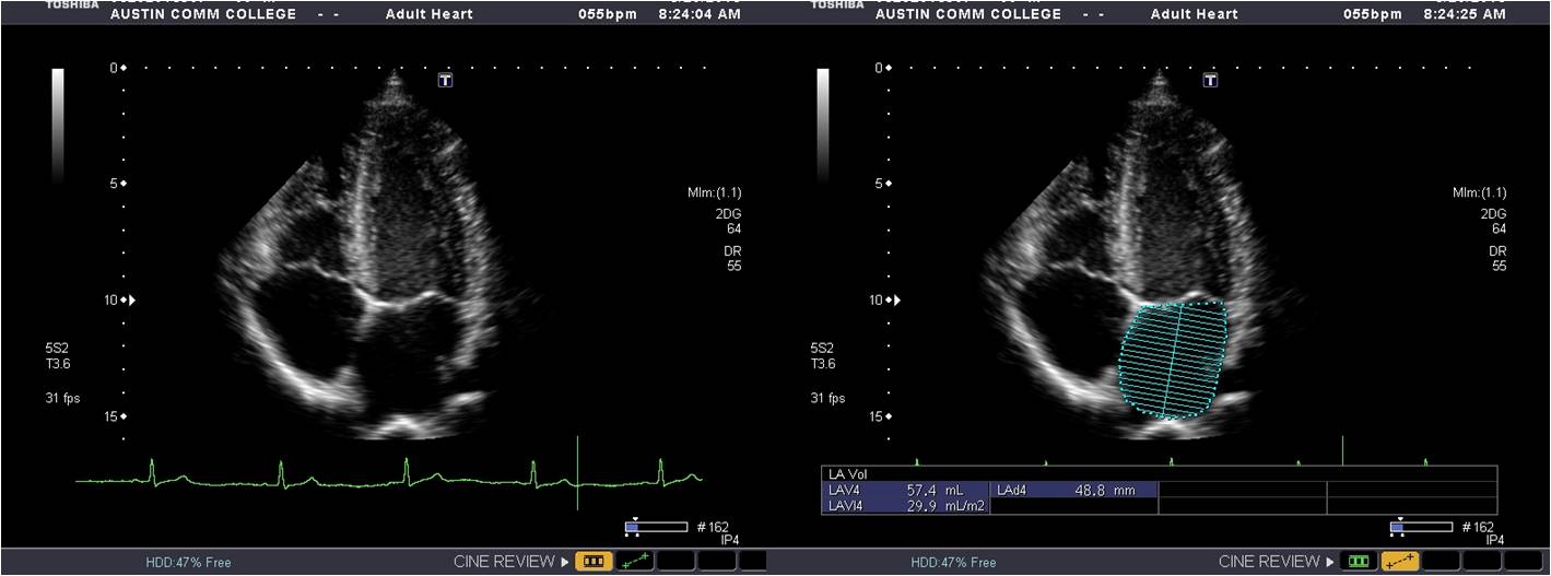
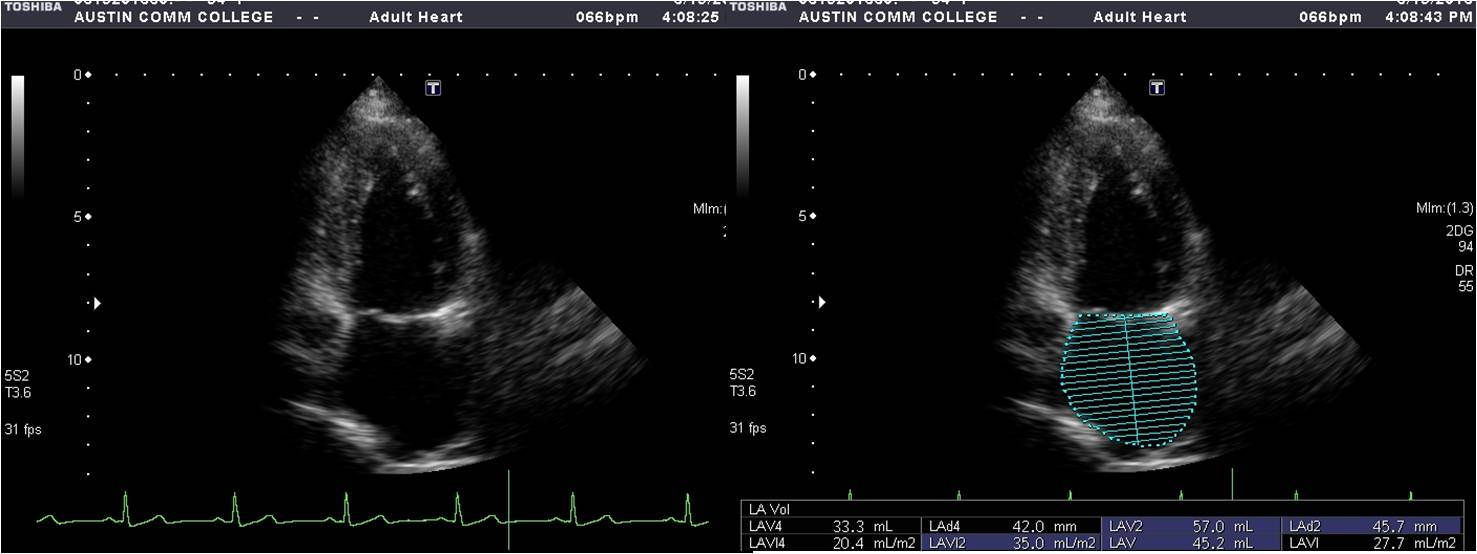
AP4C LA volume (MOD)
From an apical 4C view, optimize the LA using gains and focal zone to ensure adequate display of endocardial borders. Freeze and scroll to an end-systolic frame. Trace the borders from the anterior mitral valve annulus to the posterior mitral valve annulus. Measure the length from the center of the mitral annulus to the superior portion of the LA. Exclude the pulmonary veins and atrial appendage.
Ap2C LA volume (MOD)
From an apical 2C view, optimize the LA using gains and focal zone to ensure adequate display of endocardial borders. Freeze and scroll to an end-systolic frame. Trace the borders from the anterior mitral valve annulus to the posterior mitral valve annulus. Measure the length from the center of the mitral annulus to the superior portion of the LA. Exclude the pulmonary veins and atrial appendage.
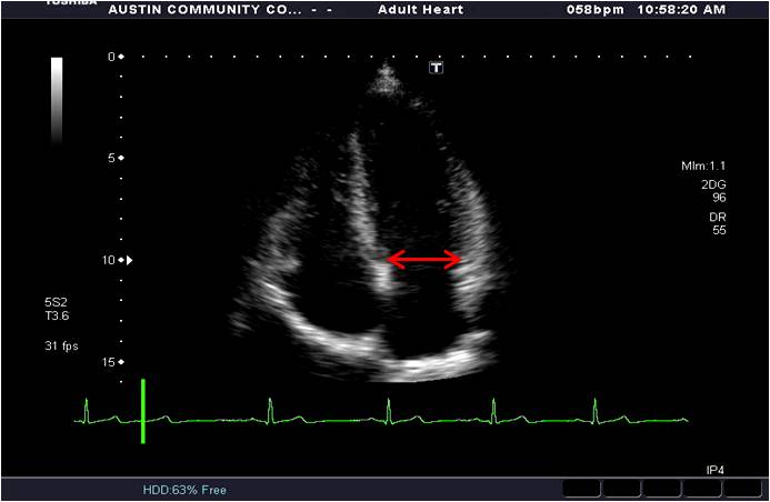
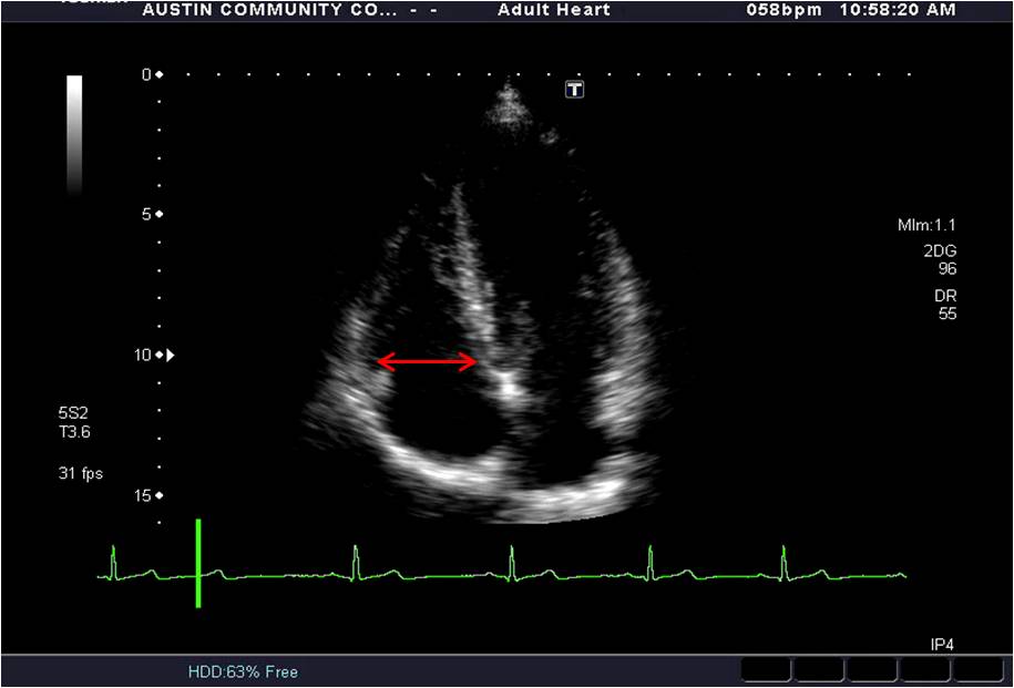
RV diameter
From an apical 4C RV focused view, freeze and scroll to an end-diastolic frame. Place the calipers at the base of the RV just distal to the tips of the tricuspid valve leaflets and on the endocardial surfaces. Ensure your measurement is perpendicular to the RV chamber.
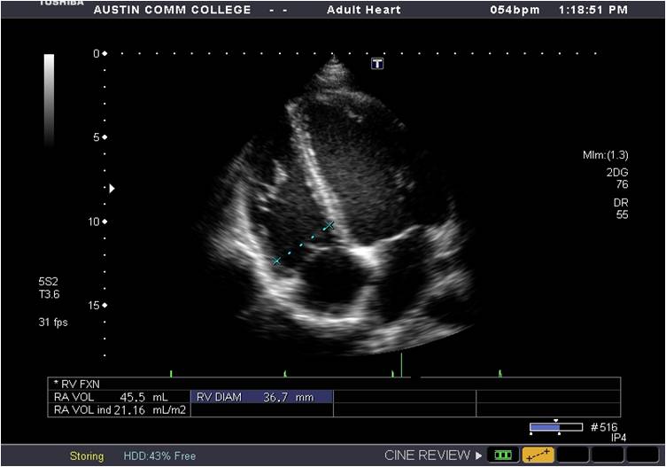
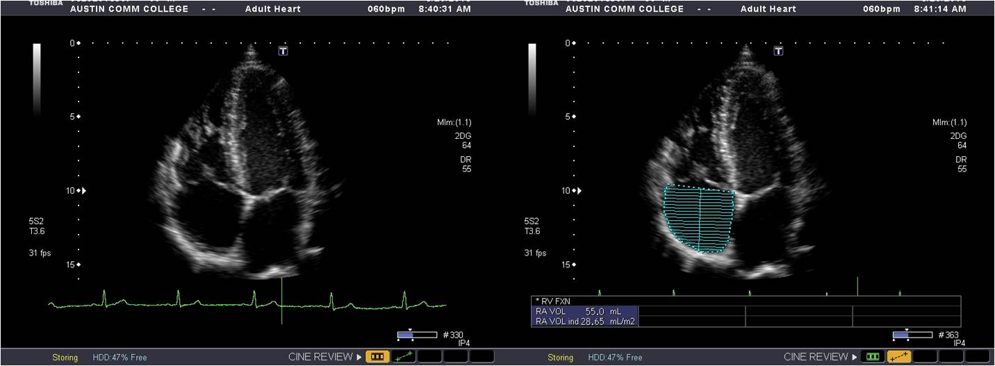
Ap4C RA volume (MOD)
From an apical 4C view, optimize the RA using gains and focal zone to ensure adequate display of endocardial borders. Freeze and scroll to an end-systolic frame. Trace the borders from the anterior tricuspid valve annulus to the septal tricuspid valve annulus. Measure the length from the center of the tricuspid valve annulus to the superior portion of the RA. Exclude the atrial appendage.