
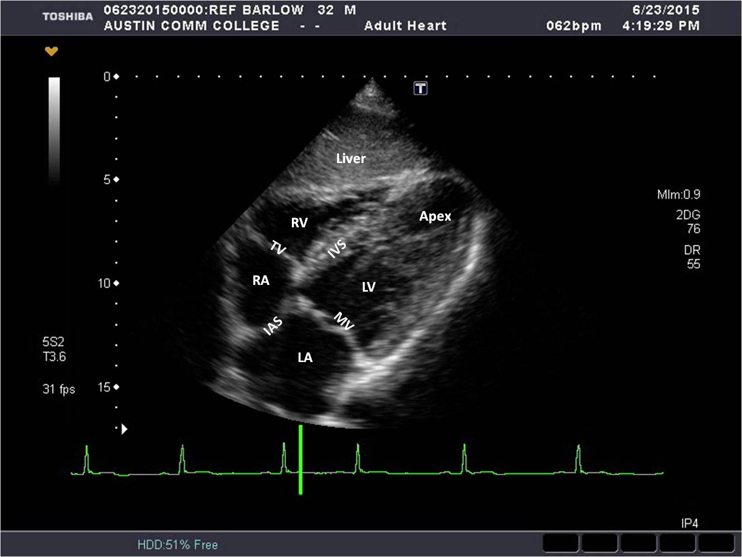
Subx 4C View
To obtain the Subxiphoid (subcostal) 4C view, place the transducer just inferior to the xiphoid process. The image marker is toward the patient’s left side and the sound beam is angled slightly anteriorly. Slight adjustments in angle and rotation maybe necessary to demonstrate all the structures for this view optimally.
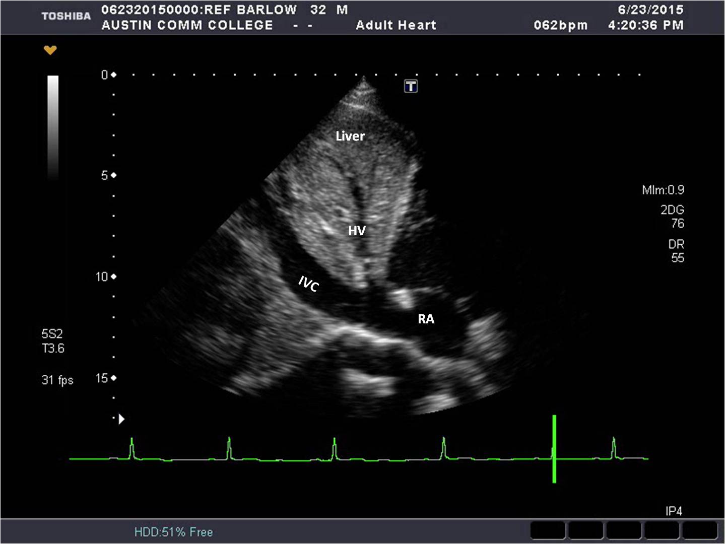
Subx @ IVC View
From the Subx 4C view, the Subx view of the IVC is obtained by rotating the transducer image marker toward the patient’s face until the IVC is elongated. Slight adjustments in angle and rotation maybe necessary to demonstrate the elongated IVC as it enters the right atrium and the hepatic vein optimally.
Subx Abd ao
From the Subx view of the IVC, the Subx view of the abdominal aorta is obtained by angling the sound beam to the patient’s left side (left lateral). Slight adjustments in angle and rotation maybe necessary to demonstrate the elongated Abd ao.

Subx SAX @ LV View
From the Subx 4C view, the Subx SAX @ LV level is obtained by rotating the transducer counterclockwise 90° until the papillary muscles are seen and the LV walls are circular. Slight adjustments in angle and rotation maybe necessary.
Subx SAX @ MV View
From the Subx SAX view @ LV, the Subx SAX @ mitral valve level is obtained by angling the sound beam toward the patient’s midline until the anterior and posterior mitral valve leaflets are seen. Slight adjustments in angle and rotation maybe necessary.
Subx SAX @ Base View
From the Subx SAX view @ MV, the Subx SAX @ the base level is obtained by angling the sound beam toward the patient’s right. Slight adjustments in angle and rotation maybe necessary.