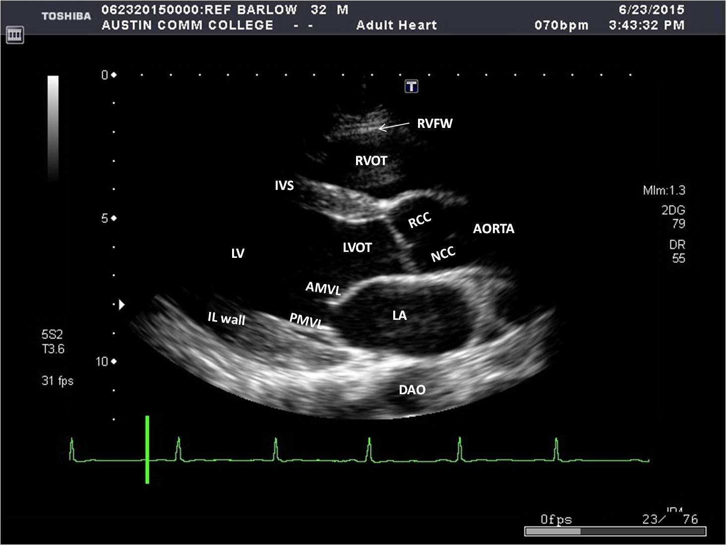
PLAX view
The parasternal long axis view (PLAX) is obtained with the transducer image marker directed toward the patient’s right ear and the sound beam directed to the spine. Slight adjustments in angle and rotation maybe necessary to demonstrate all the structures for this view optimally.
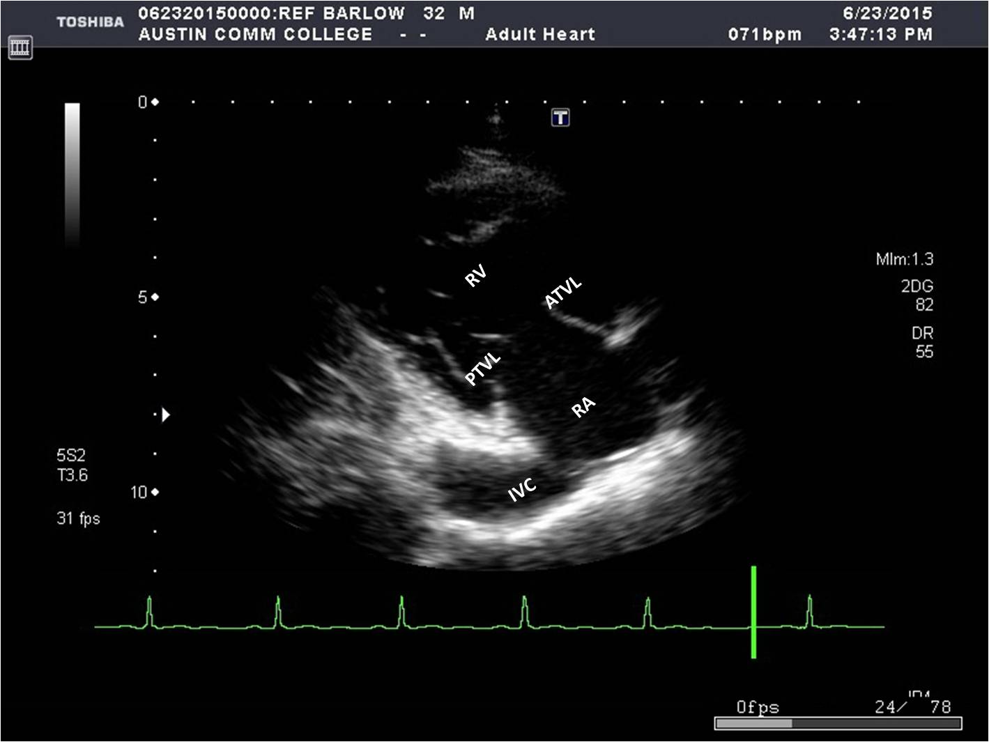
RVIT view
From the PLAX view, the right ventricular inflow tract view (RVIT) is obtained by angling the sound beam toward the patient’s right hip. Slight adjustments in angle and rotation maybe necessary to demonstrate all the structures for this view optimally.
RVOT view
From the PLAX view, the right ventricular outflow tract view (RVOT) is obtained by angling the sound beam toward the patient’s left shoulder. Slight adjustments in angle and rotation maybe necessary to demonstrate all the structures for this view optimally.
PSAX view @ base
From the PLAX view, the PSAX view @ base is obtained by rotating the transducer clockwise 90° until the image marker is directed toward the patient’s left shoulder. Slight adjustments in angle and rotation maybe necessary to demonstrate all the structures for this view optimally.

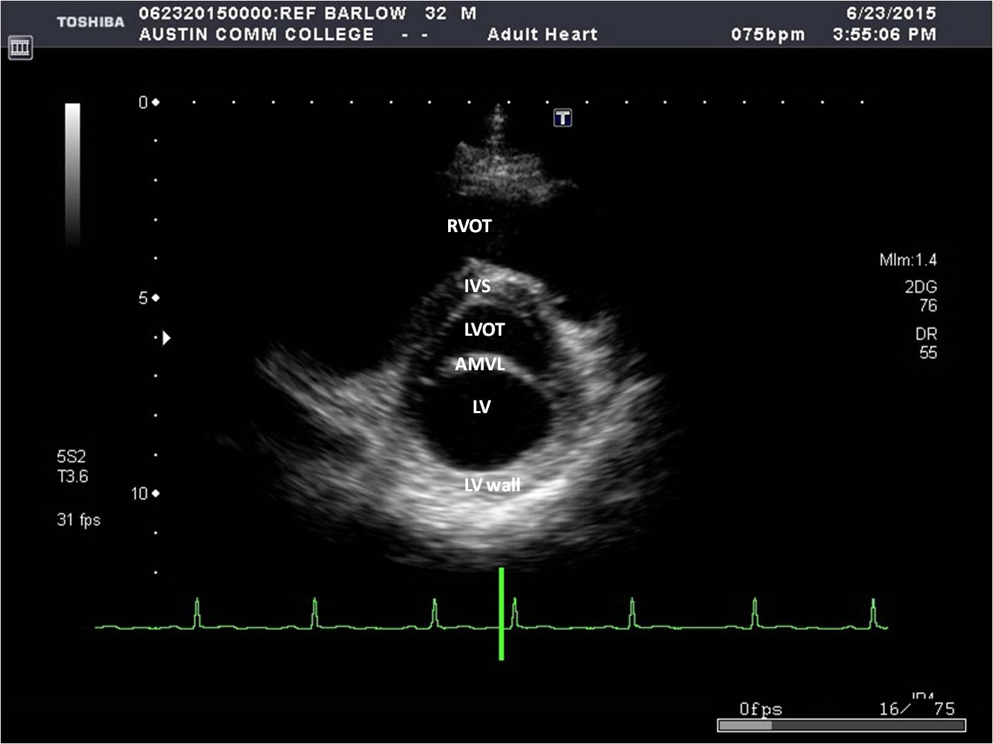
PSAX @ LVOT Level
From the PSAX view @ base, the PSAX view @ LVOT is obtained by angling the sound beam slightly inferior. Slight adjustments in angle and rotation maybe necessary to demonstrate all the structures for this view optimally.
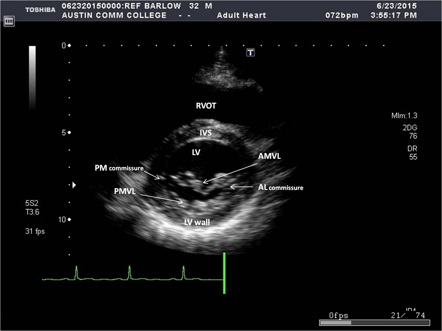
PSAX @ MV Level
From the PSAX view @ base, the PSAX view @ LVOT is obtained by angling the sound beam slightly inferior. Slight adjustments in angle and rotation maybe necessary to demonstrate all the structures for this view optimally.
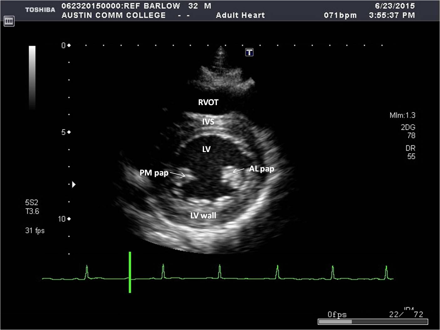
PSAX @ Pap Musc Level
From the PSAX @ MV view, the PSAX view @ papillary muscle level is obtained by angling the sound beam inferiorly until the anterior lateral and posterior medial papillary muscles are seen. Slight adjustments in angle and rotation maybe necessary to demonstrate all the structures for this view optimally.
PSAX @ apex
From the PSAX @ papillary muscle view, the LV apex level is obtained by angling the sound beam inferiorly until the LV apex is seen. Slight adjustments in angle and rotation maybe necessary to demonstrate all the structures for this view optimally.

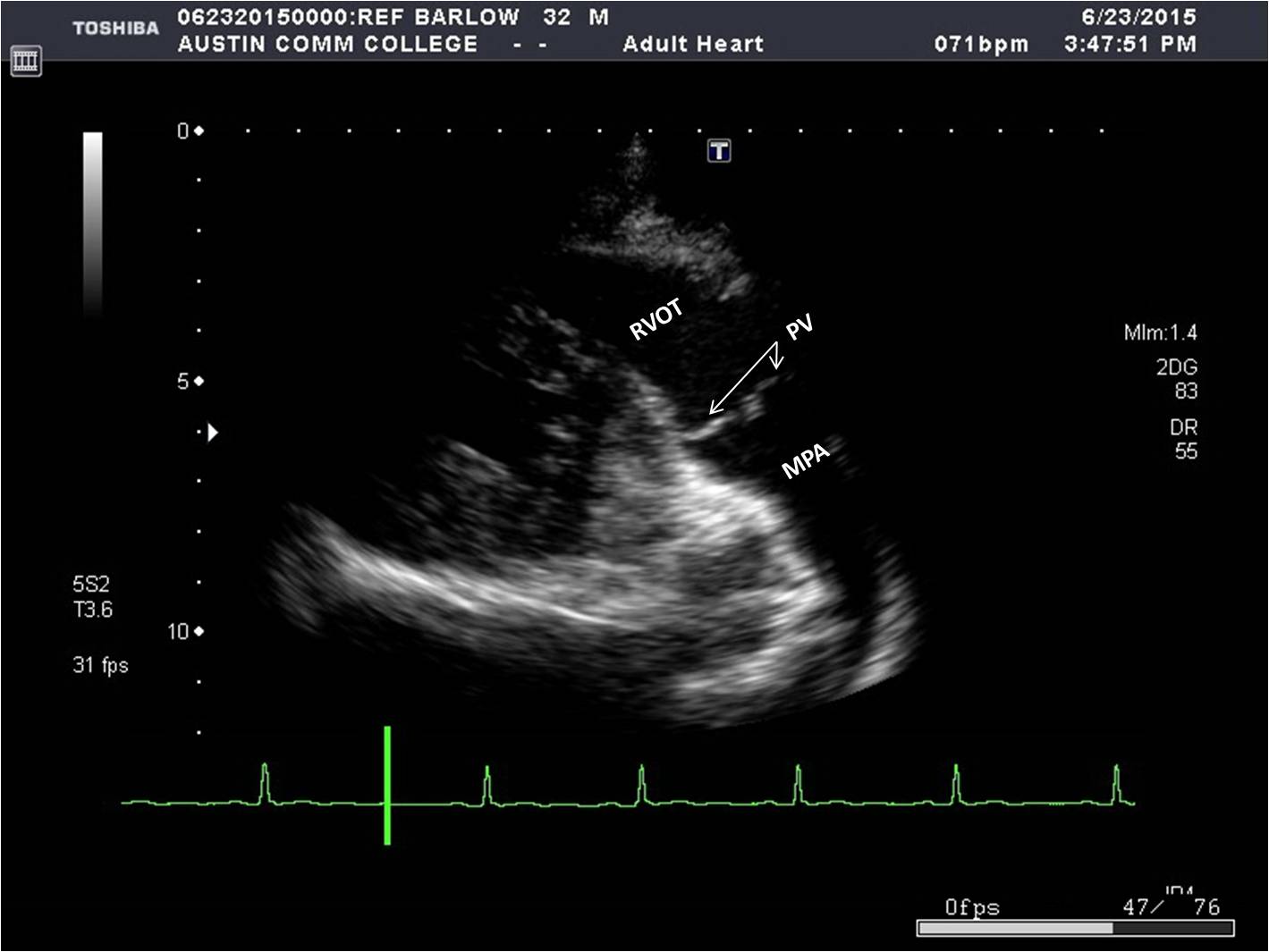
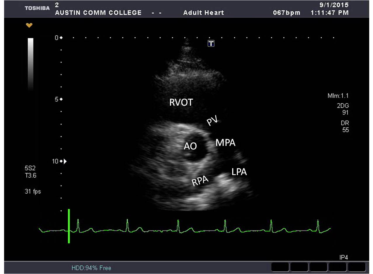
PSAX @ PA bifurc.
From the PSAX view @ base, the PSAX view @ PA bifurcation is obtained by angling the sound beam slightly more anterior and lateral until the pulmonary artery is seen. It may be necessary to use a slightly different window. Slight adjustments in angle and rotation maybe necessary to demonstrate all the structures for this view optimally.
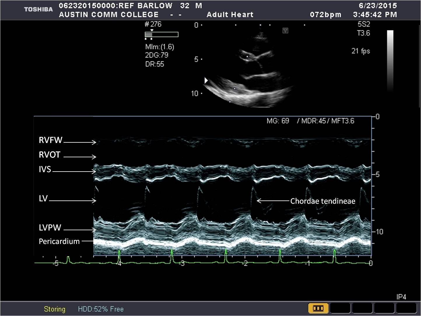
M-mode of LV
From the PLAX view, place cursor at the base of the LV, just distal to the mitral valve leaflet tips.
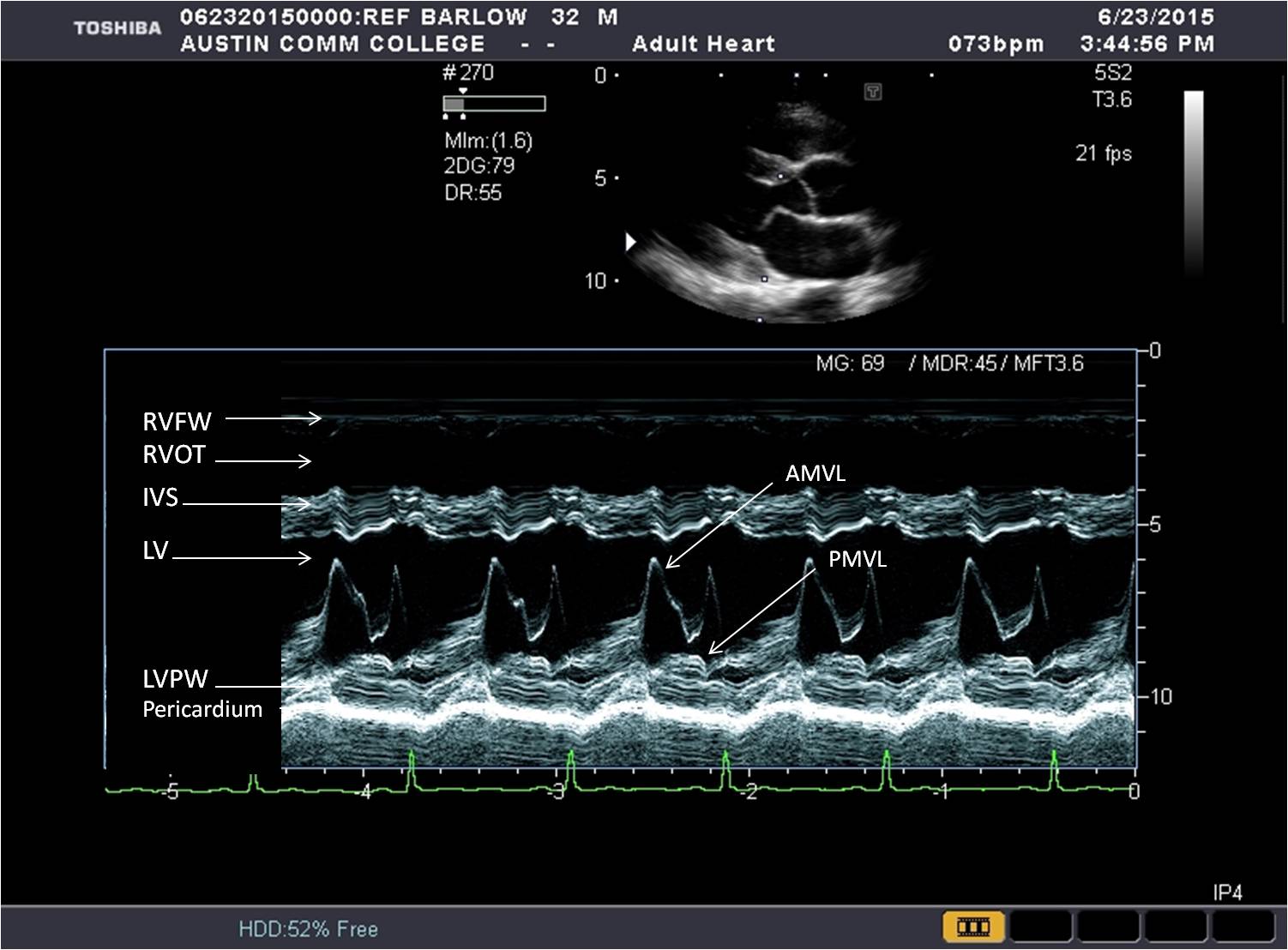
M-mode of MV
From the PLAX view, place cursor so that it transects the tips of both the anterior and posterior mitral valve leaflets.
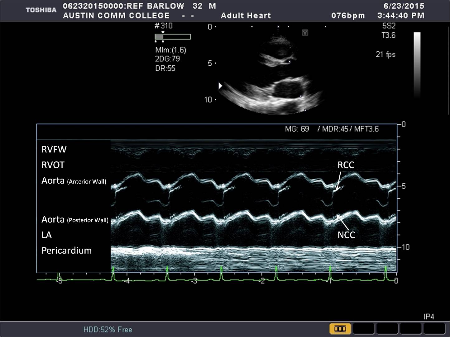
M-mode of Ao/LA
From the PLAX view, place the cursor so that it transects the closure line of the aortic valve.