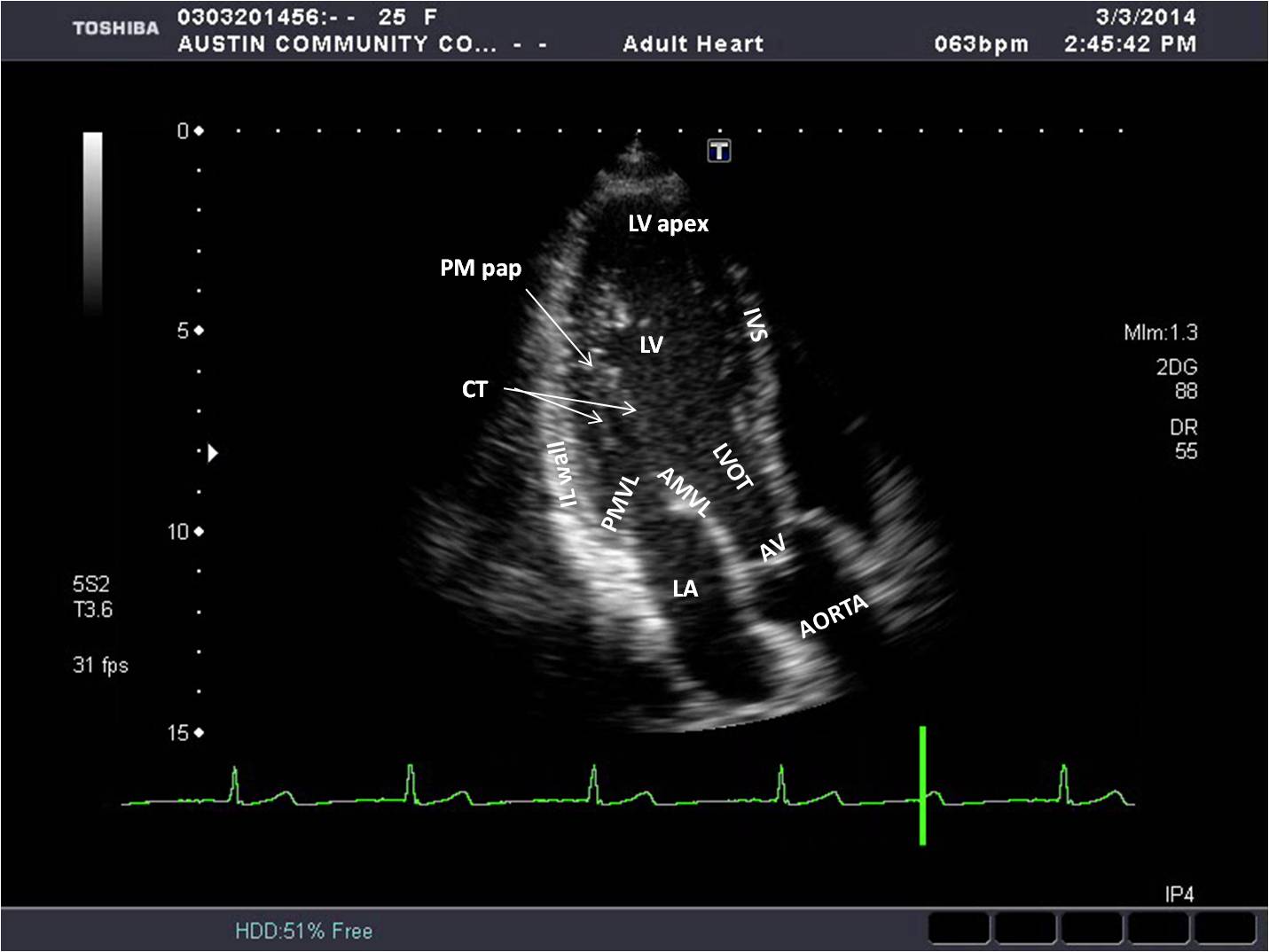
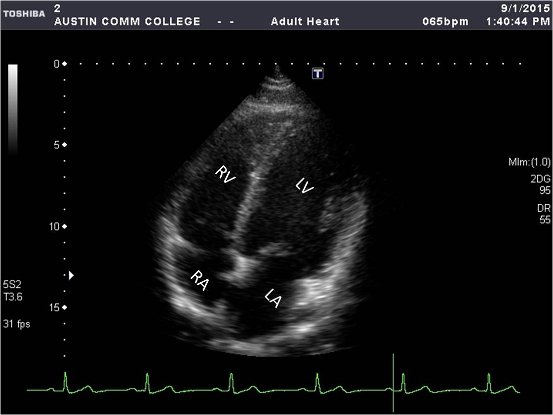
Apical 4C View
The transducer is placed at the 4-5th rib space and patient’s mid axilla with the image marker toward the bed. Ensure the sound beam transects the LV apex, all chambers are full and valves opening as widely as possible. Slight adjustments in angle and rotation maybe necessary to demonstrate all the structures for this view optimally.
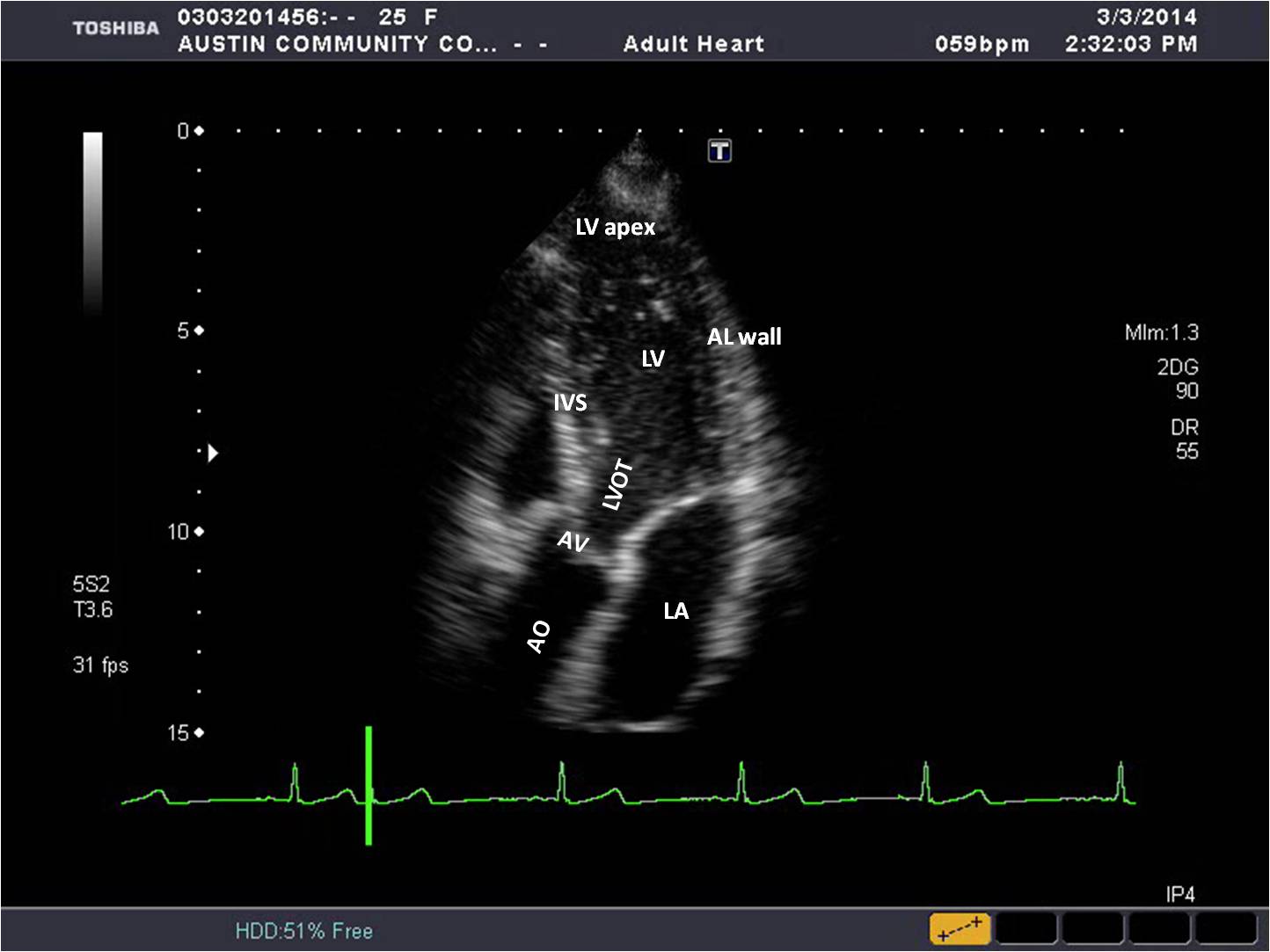
Apical 5C View
From the Apical 4C view, the Apical 5C view is obtained by angling the sound beam anteriorly and rotating clockwise until the aortic valve and aorta is seen. Slight adjustments in angle and rotation maybe necessary to demonstrate all the structures for this view optimally.
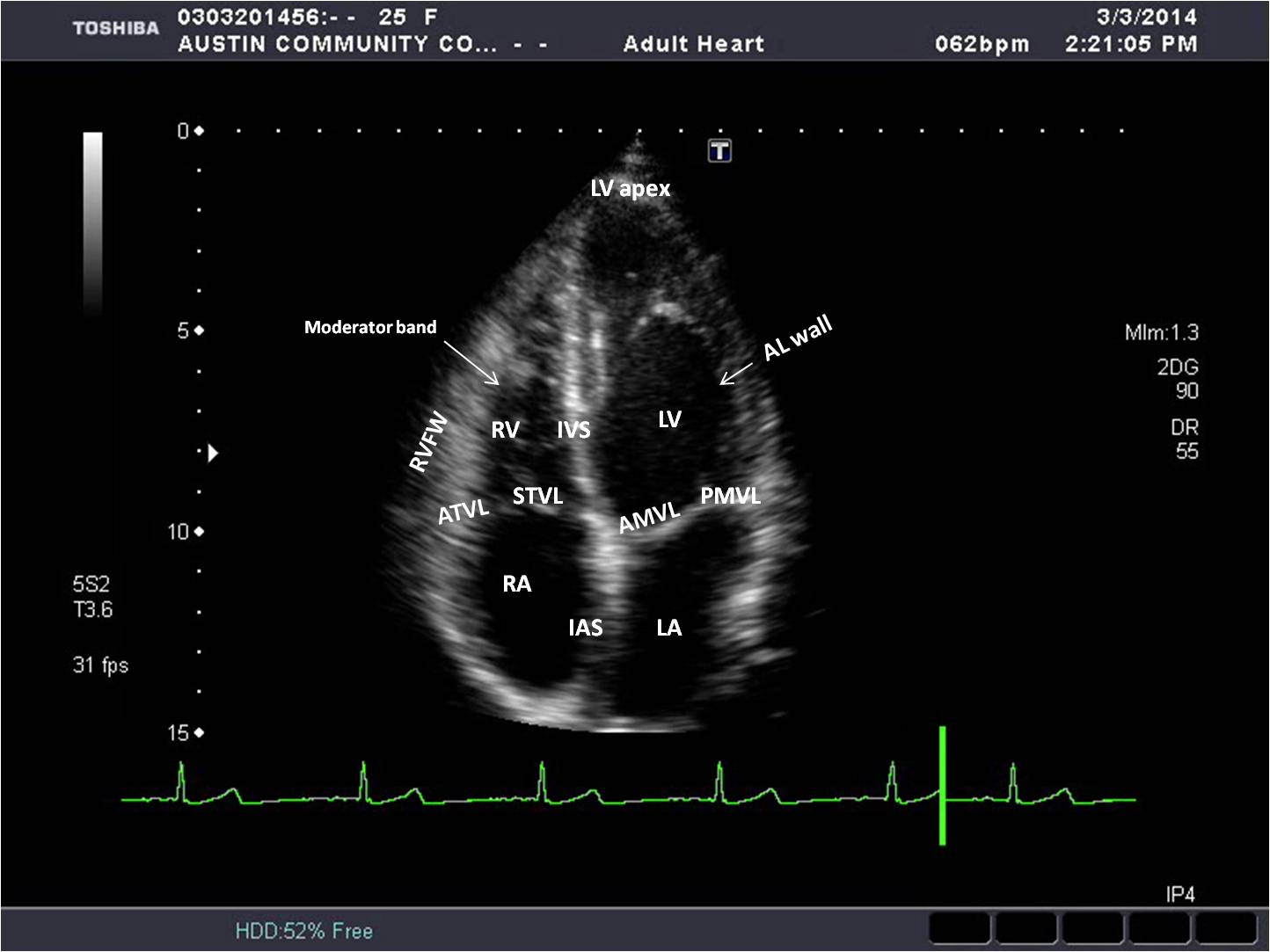
RV Focused View
From the Apical 4C view, the RV focused view is obtained by angling the sound beam medially toward the patient’s right shoulder. Slight adjustments in angle and rotation maybe necessary to demonstrate all the structures for this view optimally.
RV Modified View
From the Apical 4C view, the RV modified view is obtained by angling the sound beam laterally, toward the patient’s left shoulder. It is often necessary to move the sound beam medially as well. Slight adjustments in angle and rotation maybe needed to demonstrate all the structures for this view optimally.
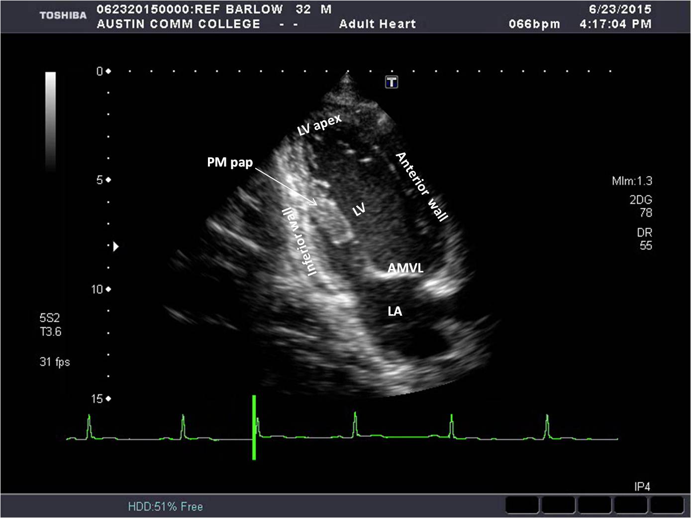
Ap 2C View
From the Apical 4C view, the Apical 2C view is obtained by rotating the sound beam 45° counterclockwise until the anterior and inferior walls are seen with careful attention to transecting the LV apex. Slight adjustments in angle and rotation maybe necessary to demonstrate all the structures for this view optimally.
Ap 3C View
From the Apical 2C view, the Apical 3C view is obtained by rotating the sound beam 45° counterclockwise until the aortic valve and aorta are fully seen. Slight adjustments in angle and rotation maybe necessary to demonstrate all the structures for this view optimally.