LV inflow @ MV tips
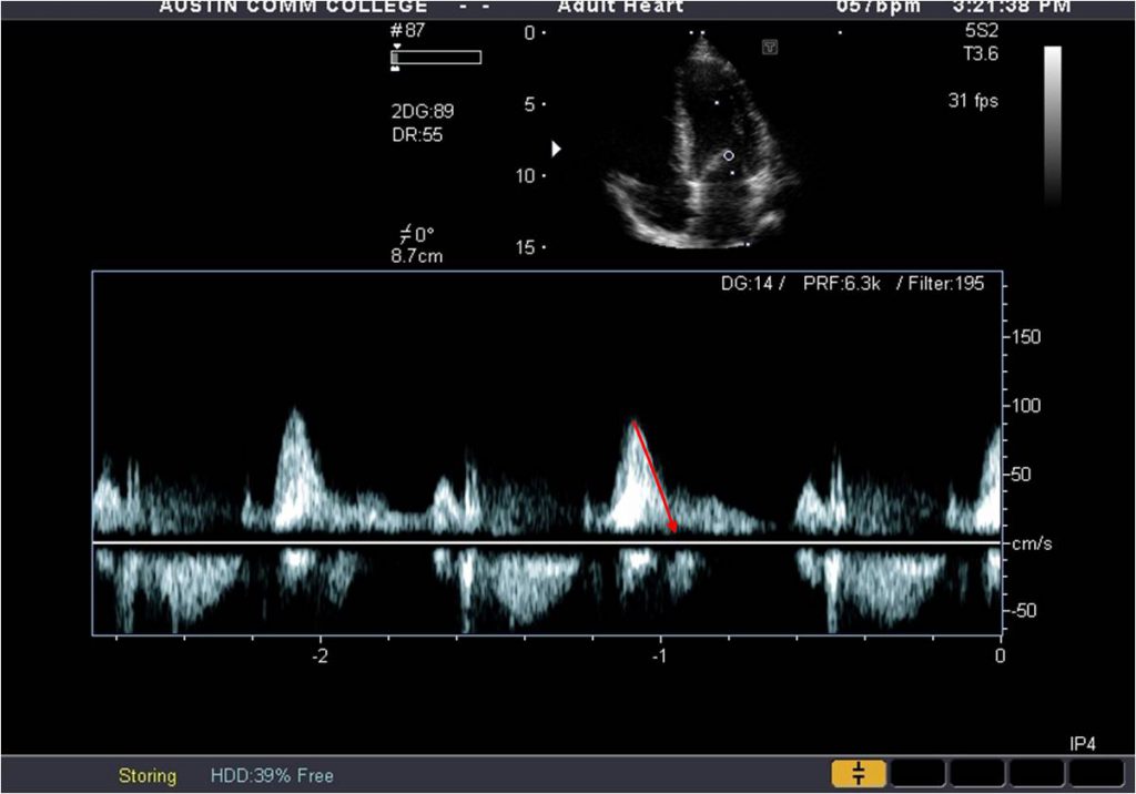
From an apical 4C view, obtain a pulsed Doppler signal of LV inflow. Place the sample volume at the tips of the mitral valve leaflets during diastole. Ensure the cursor is aligned parallel to flow of the valve. Measure the peak E, peak A and A duration.
MV VTI
From an apical 4C view, obtain a CW Doppler spectrum of the mitral flow. Place the cursor parallel to flow of the valve to ensure the highest velocities are obtained. The spectrum should demonstrate a filled-in “M” shaped spectrum. Trace the VTI.
MV PHT
From an apical 4C view, obtain a CW Doppler spectrum of the mitral flow. Place the cursor parallel to flow of the valve to ensure the highest velocities are obtained. The spectrum should demonstrate a filled-in “M” shaped spectrum. The spectrum obtained for the mitral VTI can be used. Measure the PHT by tracing the slope of the spectrum from the peak E to baseline.
Pulmonary vein measurements
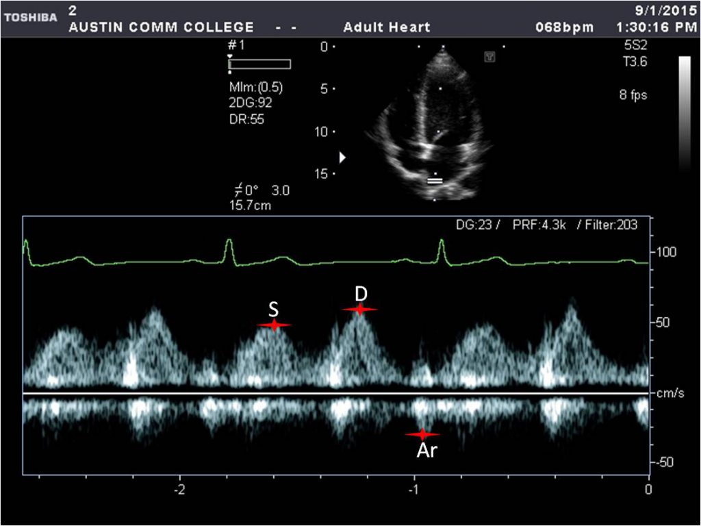
From an apical 4C view, obtain a pulsed Doppler spectrum of pulmonary venous flow. It maybe helpful to increase the depth and use color Doppler imaging to optimize the flow profile. Once the signal is obtained, measure the S wave, the D wave, and the Ar duration.
LVOT VTI
From an apical 5C view, obtain a pulsed Doppler spectrum of LV out flow. Place the sample volume just proximal to the aortic valve cusps. The spectrum should demonstrate a bright modal band with a filled in window and a closure “click”. Trace the VTI being careful to avoid the feathers around the spectrum.
AV VTI
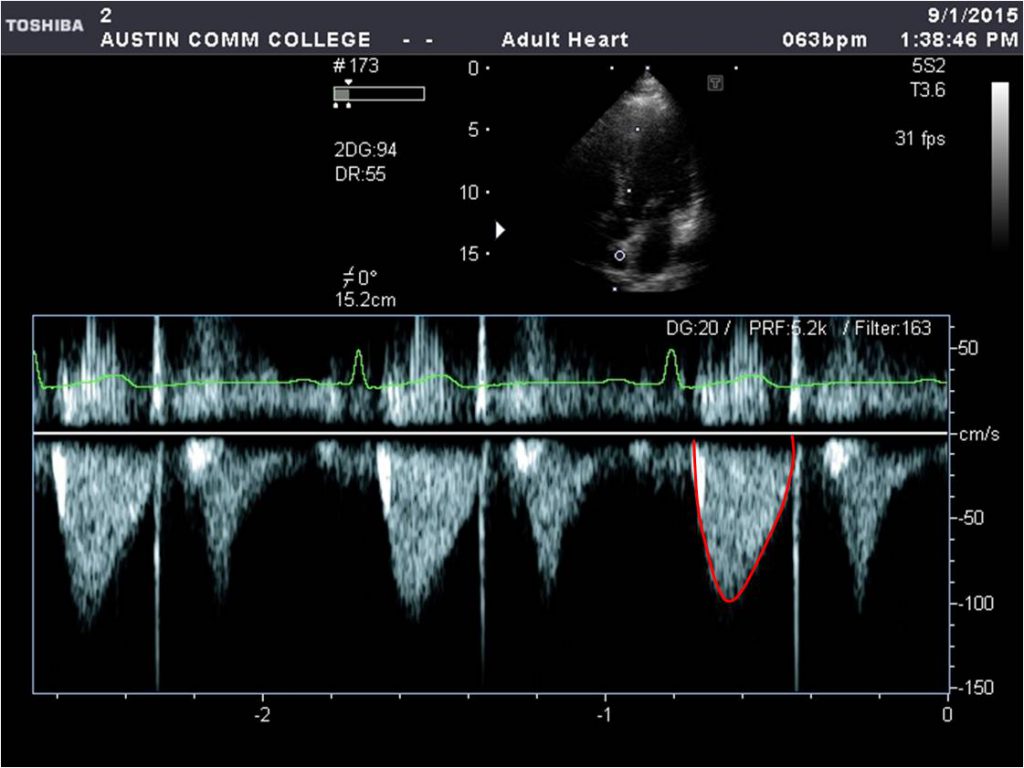
From an apical 5C view, obtain a CW Doppler spectrum of LV outflow. Place the cursor parallel to flow of this valve to ensure the highest velocities are obtained. The spectrum should demonstrate a filled-in bullet shaped spectrum. Trace the VTI careful to avoid the feathers around the spectrum.
TV VTI
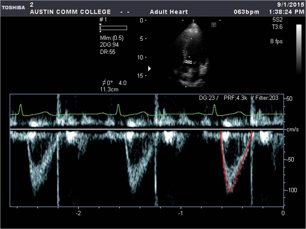
From an apical modified RV view, obtain a CW Doppler spectrum of RV inflow. Place the cursor parallel to flow of this valve to ensure the highest velocities are obtained. The spectrum should demonstrate a filled-in “M” shaped spectrum. Trace the VTI careful to avoid the feathers around the spectrum.
TDI @ septal annulus
From an apical 4C view, activate the tissue Doppler modality and align the pulsed Doppler sample volume at the septal annulus of the mitral valve. Be sure the sample volume is not on the LV or LA wall. Also, ensure the image is not angled and the LVOT is not seen.
TDI @ lateral annulus
From an apical 4C view, activate the tissue Doppler modality and align the pulsed Doppler sample volume at the lateral annulus of the mitral valve. You may need to shift the unage ti align the cursor parallel to the motion of the lateral annulus. Be sure the sample volume is not on the LV or LA wall.


