PLAX LV diameters @ end diastole
Acquire a PLAX image, freeze and scroll to an end-diastolic frame. Ensure your measurements are perpendicular to the structures and chambers. Place the caliper on the surface of the endocardial borders.

PLAX LV diameter @ end systole
Acquire a PLAX image, freeze and scroll to an end-systolic frame. Ensure your measurements are perpendicular to the structures and chambers. Place the caliper on the surface of the endocardial borders.

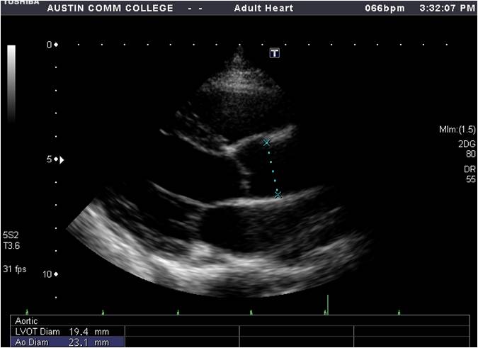
PLAX Ao diameter
From a PLAX image, freeze and scroll to an end-diastolic frame. Measure at the sinotubular junction using the inner edge to inner edge technique
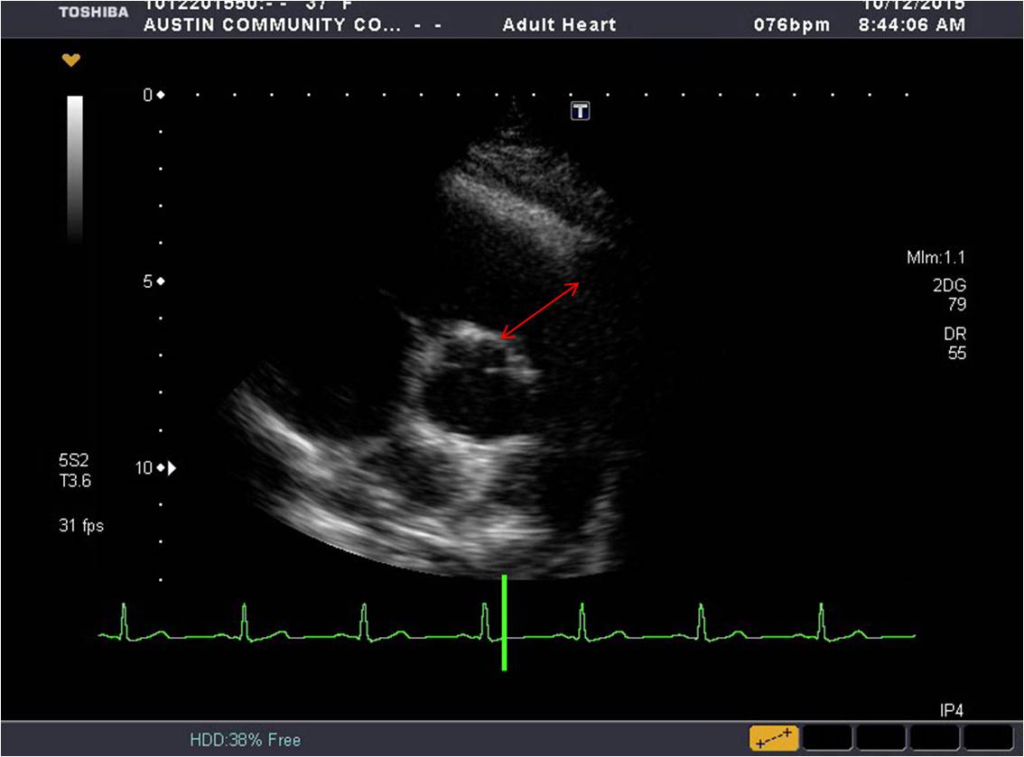
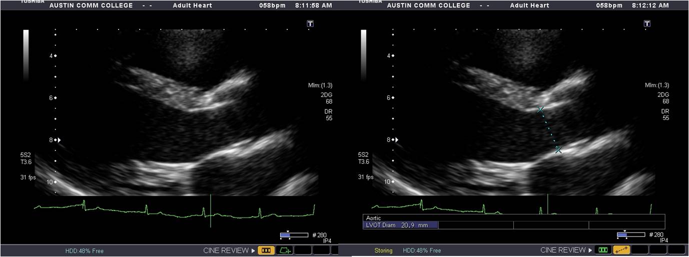
PLAX LVOT diameter
Acquire a PLAX image, ZOOM the aortic valve and freeze. Scroll to an early systolic frame ensuring the cusps and the insertions of the aortic valve are displayed. Measure at the insertions.
PSAX RVOT diameter
From a PSAX at the base, obtain optimal images of the pulmonic valve and the insertion of the cusps into the artery. Freeze and scroll to early to mid systole (the insertions should be clearly seen). Measure from where the right anterior cusp inserts into the annulus to the left anterior cusp insertion using the inner edge to inner edge technique.