
PW of LV inflow (tips)
From the apical 4C view, place the PW Doppler sample volume at the tips of the mitral leaflets. Optimize the signal to eliminate spikes and feathering.
PW of LV inflow (annulus)
From the apical 4C view, place the PW Doppler sample volume at the annulus of the mitral valve. Optimize the signal to eliminate spikes and feathering.
CW of LV inflow
From the apical 4C view, align the cursor across the center of mitral valve flow. Optimize the signal to eliminate spikes and feathering.
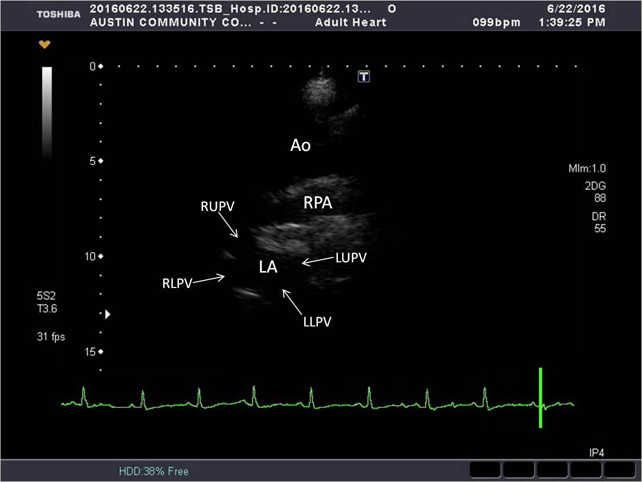
PW of Pulm Venous Flow
From the apical 4C view, optimize the left atrium by increasing the depth. Using color Doppler, angle and rotate to optimize flow of the right upper pulmonary vein. Place the PW Doppler sample volume in the flow and activate Doppler.
PW LVOT
From the apical 5C view, place the PW sample volume just proximal to the aortic valve cusps. Optimize the signal to eliminate spikes and feathering.
CW AV
From the apical 5C view, align the cursor center of aortic valve flow. Optimize the signal to eliminate spikes and feathering.
CW TV
From the apical 4C modified RV view, align the CW Doppler cursor center of tricuspid valve flow. Optimize the signal to eliminate spikes and feathering.
[QTL-Question id=21]
[QTL-Question id=22]
[QTL-Question id=23]